
Radiation Shielding Update
A New Era of Dental Imaging Safety in Missouri
by Stephanie Patrick, BS,RT(R)(M)(QM)
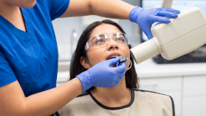
Are we hanging up the lead apron for good? In recent years, the American Dental Association and the American Academy of Oral and Maxillofacial Radiology have recommended that dental offices discontinue the use of lead aprons and thyroid shields for patients, regardless of age.1,2 There have been some conflicting recommendations regarding patient shielding, as the Missouri Radiation Control Program (MRCP) follows the National Council on Radiation Protection and Measurements (NCRP) guidelines for radiation protection, rather than those of the dental organizations.
Over the last few years, the NCRP has had a scientific committee comprised of radiation safety experts review the long-standing recommendations for shielding for all radiology exams and modalities, including dental imaging. A recent publication, NCRP Commentary No. 35: Patient Shielding in Medical Imaging, was released on July 1, 2026.
The NCRP made the following statement: “Shielding is not recommended for any anatomy during dental and maxillofacial imaging, including intraoral radiographs, panoramic and cephalometric imaging, and CBCT. This recommendation is for all patients regardless of age or sex.”3 This statement is an updated recommendation from the previous NCRP Report No. 177, where thyroid shielding was recommended during dental imaging.4
The MRCP has confirmed in written correspondence with the author that it will adhere to the NCRP recommendations in Commentary No. 35. Consistent with those recommendations, routine patient shielding is not recommended; however, the MRCP has stated that it will not actively discourage facilities from providing leaded PPE when a facility believes it may provide psychological comfort to patients, as this has become the norm in many offices.
Why did the recommendation change?
Modern digital receptors and optimized dental imaging techniques allow diagnostic images to be obtained with substantially lower radiation exposures than with older technologies.1-3 In addition, because radiation-induced heritable effects have not been demonstrated in humans, the scientific rationale for routine gonadal shielding has changed.1-3 The NCRP also notes that thyroid radiosensitivity decreases with age, and a 2023 systematic review found no strong evidence of an increased thyroid cancer risk from radiation exposure during adulthood.3,5
Since dental imaging now uses less radiation, the amount of internal scatter produced has also decreased. As primary beam photons from the x-ray tube enter the patient’s body tissues and create internal scatter, they can interact with the lead shield and create additional scattered photons (backscatter to the patient). The primary beam itself is collimated and does not directly contribute to radiation dose to organs outside the field of view, such as the gonads; dose to these organs is primarily attributable to internal scattered radiation.3,6 In addition, the larger the patient, the greater the amount of internal scatter produced, as there is more tissue in the body with which the x-rays can interact. Patient shielding does not decrease dose from internal scatter.
When a panoramic or cone beam CT image is taken, the x-ray beam is moving completely around the patient, so if a patient is wearing a thyroid shield, the only time the thyroid is blocked from radiation is when the beam is briefly in front of the patient. As the beam moves, the thyroid shield offers no additional protection.3 Dose to the thyroid may increase in this situation due to back-scattered radiation from the thyroid shield, although this effect is minimal when using a shield with high atomic number materials, such as lead.3
In addition, thyroid shields can block the anatomy of interest depending on the neck length of the patient or in children unable to hold still for imaging; artifacts may also result if the shield is positioned closely to the mandible, increasing the likelihood that a repeat image will be required.1-3
When performing dental imaging on pregnant patients, the fetus is outside of the imaging area. Any radiation dose to the fetus will be from internal scattered radiation within the mother; external shielding will not decrease dose to the fetus, so a lead apron should not be utilized.7,8 Shielding a fetus may not provide radiation protection, but dental staff may encounter patients who feel reassured by a lead apron.3
What are the best practices to reduce the radiation dose to the patient as much as possible?
Reducing dose to the patient can be accomplished by precise positioning to avoid repeated images, using a lower mA and time setting (mAs), and utilizing rectangular collimation at the end of the cone during intraoral radiographs. Rectangular collimation is not widely utilized but can decrease patient dose by 40% or more compared with circular collimation.3,9,10 All dental x-ray units have adjustable exposure selections, and the appropriate settings should be selected according to the patient’s size. Utilizing these factors is more beneficial to the patient than shielding the thyroid.
Patients directly benefit from imaging to determine treatment based on recommendations from their dental provider. Dental personnel who are occupationally exposed to radiation do not receive that direct imaging benefit and should continue to follow occupational radiation-protection practices, including maintaining distance and using structural shielding and remote exposure switches where applicable.3
In summary, the NCRP no longer recommends the use of thyroid shields or lead aprons for any patients undergoing dental imaging; even if the thyroid area is included in imaging, a shield is not recommended. Radiation dose to organs outside the imaging field, including the thyroid and gonads, results primarily from internal scatter in the patient, and external shielding will not assist in decreasing radiation dose to these organs. Using low-exposure techniques, accurate positioning, and rectangular collimation on cones are the most effective ways to decrease radiation dose to the patient.
The MRCP has confirmed that it will adhere to the NCRP recommendations in Commentary No. 35 with the caveat that it will not actively discourage facilities from providing leaded PPE when a facility believes it provides psychological comfort to patients.
 Stephanie Patrick is a Qualified Expert with Heldebrandt Consulting and travels throughout Missouri performing radiation safety inspections for sites including dental settings. She has been in the radiologic sciences with experience in radiation physics and safety for more than 30 years and enjoys providing educational radiology information to facilities. Contact Stephanie for questions or to learn more about inspections.
Visit the MDA Radiation page for complete information on inspections, a link to the MRCP survey schedule for dental practices, various FAQ and more.
FAQ & Answers from Stephanie Patrick
How often does my x-ray equipment have to be inspected? Does every dental facility have to be surveyed?
The MRCP assigns a class D or E to dental sites depending on the type of equipment that is located and in use at that site. Class D equipment includes panoramic units (2D units), intraoral wall units, and handheld units; these units are due for surveying every six years. Class E equipment is for cone beam CT units (3D units), and those x-ray units specifically are on a three year survey cycle because they produce more radiation. Yes, every site is mandated to have the units tested to remain in compliance with the MRCP.
I only have Nomads. Do these units have to be surveyed?
Yes. Any equipment that produces ionizing radiation at your office must be registered with the MRCP and surveyed by a qualified expert.
My equipment is due for inspection at the end of the year. If it is surveyed earlier, does that change the due date?
No, your due date remains the same. Equipment can be surveyed up to 364 days in advance of your due date and be acceptable to the MRCP. Early is great, late will result in a warning letter from the MRCP, which we like to avoid.
I am moving my office. Does everything have to be inspected again?
Yes. Whether you are bringing your previous equipment to your new site or purchasing new equipment (or a combination), all equipment at the new location must be inspected. This new survey date will start a new survey cycle. You will keep the same facility ID that is registered with the MRCP. If any x-ray equipment is in storage or removed by a vendor, that must be noted on your registration form as well.
References
- Benavides E, Bhula A, Gohel A, Lurie AG, Mallya SM, Ramesh A, et al. Patient shielding during dentomaxillofacial radiography: recommendations from the American Academy of Oral and Maxillofacial Radiology. J Am Dent Assoc. 2023;154(9):826-835.e2. doi:10.1016/j.adaj.2023.06.015.
- Benavides E, Krecioch JR, Connolly RT, Allareddy T, Buchanan A, Spelic D, et al. Optimizing radiation safety in dentistry: Clinical recommendations and regulatory considerations. J Am Dent Assoc. 2024;155(4):280-293.e4. doi:10.1016/j.adaj.2023.12.002.
- National Council on Radiation Protection and Measurements. Patient shielding in medical imaging. NCRP Commentary No. 35. Bethesda (MD): National Council on Radiation Protection and Measurements; 2026.
- National Council on Radiation Protection and Measurements. Radiation protection in dentistry and oral & maxillofacial imaging: recommendations of the National Council on Radiation Protection and Measurements. NCRP Report No. 177. Bethesda (MD): National Council on Radiation Protection and Measurements; 2019.
- Mirkatouli NB, Hirota S, Yoshinaga S. Thyroid cancer risk after radiation exposure in adults—systematic review and meta-analysis. J Radiat Res. 2023;64(6):893-903. doi:10.1093/jrr/rrad073.
- Matyagin YV, Collins PJ. Effectiveness of abdominal shields in chest radiography: a Monte Carlo evaluation. Br J Radiol. 2016;89(1066):20160465. doi:10.1259/bjr.20160465.
- Kelaranta A, Ekholm M, Toroi P, Kortesniemi M. Radiation exposure to foetus and breasts from dental X-ray examinations: effect of lead shields. Dentomaxillofac Radiol. 2016;45(1):20150095. doi:10.1259/dmfr.20150095.
- American College of Radiology, Society for Pediatric Radiology. ACR-SPR practice parameter for imaging pregnant or potentially pregnant patients with ionizing radiation. Revised 2023. Reston (VA): American College of Radiology; 2023.
- Johnson KB, Ludlow JB. Intraoral radiographs: a comparison of dose and risk reduction with collimation and thyroid shielding. J Am Dent Assoc. 2020;151(10):726-734. doi:10.1016/j.adaj.2020.06.019.
- Shetty A, Almeida FT, Ganatra S, Senior A, Pacheco-Pereira C. Evidence on radiation dose reduction using rectangular collimation: a systematic review. Int Dent J. 2019;69(2):84-97. doi:10.1111/idj.12411.
